
I had asked him to see if he could get a printout of the CT reports from both his pre-surgery CT scan and his most recent CT scan. I figured if I had something to read and flip through, I might be able to understand it better. And whatever I didn't understand, I knew I could ask any of the numerous medical people I work with to help explain some of the unfamiliar terminology.
Below are some passages from the reports (C1 - 1st CT scan done pre-surgery, and C2 - 2nd CT scan done post-chemo) with translations and pictures.
C1
Liver mildly enlarged, measuring 17 cm in length, with subcentimeter hypodensity lesion in the right hepatic lob too small to accurately characterize.
--translation: The liver is mildly enlarged with a very tiny (less than a centimeter) lesion (dark mass) on the right hepatic lobe that is so small it cannot be characterized as anything.
2 enlarged hypervascular left para-aortic lymph nodes measuring 1.7x2.0 and 1.9x2.1 cm.
--translation: 2 lymph nodes on the aorta are enlarged to approximately 2 cm.
Impression (translation: summary)
Left para-aortic nodal metastases. No other findings of metastatic disease in the chest, abdomen, and pelvis. Mild hepatomegaly with subcentimeter hypodensity too small to accurately characterize; however, it is favored to represent a tiny cyst.
--translation: Cancer has metastasized (spread) to the para-aortic lymph nodes (lymph nodes around the aorta), but not anywhere else. The liver is swollen a little and the lesion on the liver is suspected to be a tiny cyst. (Note: The aorta is not just an artery that surrounds the heart, but it also extends down into the main body cavity....so "para-aortic lymph nodes" are not, in fact, anywhere near the heart. Just FYI.)
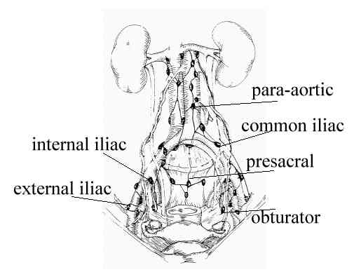 |
| para-aortic lymph node group, just under the LEFT kidney |
Other interesting things noted in C1:
Small fat-containing umbilical and left inguinal hernias
--translation: some small internal hernias, but nothing to worry about
Minimal degenerative changes of the thoracolumbar spine
--translation: he's shrinking in stature
Mild bilateral gynecomastia
--translation: swelling of the breast tissue (upon further research, I learned that this is common with hormonal imbalances due to testicular cancer)
And the best phrase that I'm sure he's going to love: extratesticular enhancement
C2
Within the abdomen, there is normal appearance of the liver, gallbladder, pancreas, bilateral adrenal glands and bilateral kidneys.
--translation: the liver and all subsequent area organs look normal (no mention of previous swollen liver or lesion)
There are enhancing and enlarged retroperitoneal lymph nodes at the level of the left kidney measuring 1.5 cm and 1.8 cm in diameter, relatively unchanged since the previous exam. There is no new lymphadenopathy.
--translation: 2 lymph nodes in the retroperitoneal space by the left kidney are measuring 1.5 cm and 1.8 cm and are thus only a little smaller than the time of his last scan. There are, however, no NEW enlarged lymph nodes (in the abdominal area)
Impression (translation: summary)
Stable retroperitoneal lympadenopathy. No new lymph node enlargement. Mild gynecomastia.
--translation: lymph nodes that were there before are still there, but there aren't any new nodes. Breast tissue still swollen (further research said it would take a while for his hormones to balance out after surgery and may result in continued swelling.)
.jpg) |
| This picture depicts the entire "retroperitoneal space"....note that the aorta (in red) and para-aortic lymph nodes (nodes are green) are included |
So the confusion with the results last Friday was this:
One doctor kept saying there was no mention of the aortic lymph nodes, but there was an area observed by the left kidney that was most likely calcification. The other doctor kept talking about the retroperitoneal nodes that were less swollen than before and more testing was needed. But nobody told us that the "para-aortic lymph nodes" and "retroperitoneal lymph nodes behind the left kidney" were the SAME lymph nodes. Turns out...the aorta is located within the retroperitoneal space...and the lymph node group called "para-aortic" is located just under/behind the left kidney.
Definition of the retroperitoneal space
"The retroperitoneal space is bounded by the posterior parietal peritoneum anteriorly and the lumbar spine posteriorly. The retroperitoneal space contains the kidneys, adrenal glands, pancreas, nerve roots, lymph nodes, abdominal aorta, and inferior vena cava. The retroperitoneal (or lumbar) lymph nodes are the regional lymph nodes for the organs of the retroperitoneal space, and also for the testes, ovaries, fallopian tubes, and uterus (which are embryologically derived from the retroperitoneum). The retroperitoneal nodes are divided by the aorta and inferior vena cava into three groups: those lying to the left of the aorta (left para-aortic or left lumbar group), those lying between the aorta and inferior vena cava (interaortocaval or intermediate lumbar group), and those lying to the right of the inferior vena cava (right paracaval or right lumbar group)."
After having been able to read the reports myself and have the terminology explained by other medical people who AREN'T oncologists, we have learned that the two lymph nodes that were swollen to about 2 cm each before the chemo are, in fact, still swollen, but have reduced minimally in size. We were told that, with seminomas, its possible for lymph nodes to remain swollen even after all the cancer cells have been killed. The PET scan he had yesterday will determine if there are any more cancerous cells alive and kickin'. The tumor board will discuss his case and decide if his lymph nodes still might have some residual seminoma cells, or if they're a teratoma, or if they are just swollen.
(Fun fact: A teratoma is one of those tumors that is often seen on TV when they tell a person that they were a twin in utero and their body absorbed the other twin. But that's not just what a teratoma is. A teratoma is a tumor that is made up of all kinds of extraneous tissue cells without having to have absorbed it from a fetus. A teratoma may be found to contain multiple types of tissue cells, including specialize organ cells, bone, hair, etc.)
Lessons learned: always get a copy of the report. Even though you're not a doctor, you may be able to understand the report better than if a doctor is standing in front of you and all of a sudden throwing out words like "kidney...teratoma...lymphoma..." Those scary words aren't necessary until AFTER the reports have been looked at.
I don't know about Dave...but I'm a little less stressed knowing what I learned today.
No comments:
Post a Comment